Home

Talla Acusación puño raman cell micro Pronombre Excretar

Visualizing Cell State Transition Using Raman Spectroscopy | PLOS ONE

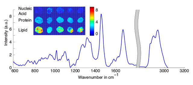
Single cell Raman spectroscopy for cell sorting and imaging - ScienceDirect

Raman spectroscopy for physiological investigations of tissues and cells -  ScienceDirect
Raman spectroscopy for physiological investigations of tissues and cells - ScienceDirect

Raman microscopy for cellular investigations — From single cell imaging to  drug carrier uptake visualization - ScienceDirect
Raman microscopy for cellular investigations — From single cell imaging to drug carrier uptake visualization - ScienceDirect

Visualizing Cell State Transition Using Raman Spectroscopy | PLOS ONE

Non-invasive cell classification using the Paint Raman Express Spectroscopy  System (PRESS) | Scientific Reports
Non-invasive cell classification using the Paint Raman Express Spectroscopy System (PRESS) | Scientific Reports

The substrate matters in the Raman spectroscopy analysis of cells |  Scientific Reports
The substrate matters in the Raman spectroscopy analysis of cells | Scientific Reports

Single cell Raman spectroscopy for cell sorting and imaging - ScienceDirect

Sensors | Free Full-Text | Raman Spectroscopy Cell-based Biosensors

Biomolecular phenotyping and heterogeneity assessment of mesenchymal  stromal cells using label-free Raman spectroscopy | Scientific Reports
Biomolecular phenotyping and heterogeneity assessment of mesenchymal stromal cells using label-free Raman spectroscopy | Scientific Reports

Using Raman Spectroscopy to Identify Cell Types | The Scientist Magazine®

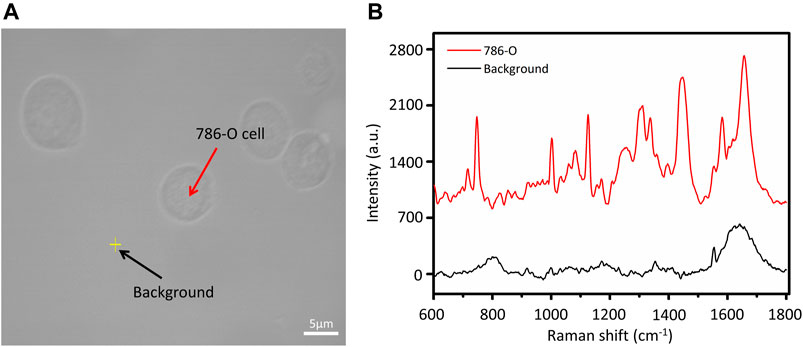
Frontiers | Detection and Classification of Multi-Type Cells by Using  Confocal Raman Spectroscopy
Frontiers | Detection and Classification of Multi-Type Cells by Using Confocal Raman Spectroscopy

Raman spectroscopy for label free cells analysis | LabionLabion

Raman Electrochemical Flow Cell Setup– MSE Supplies LLC

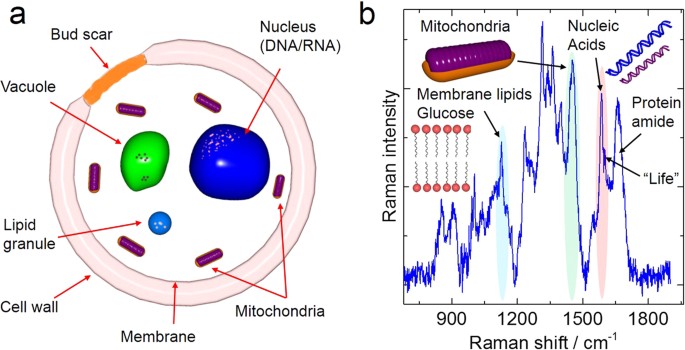
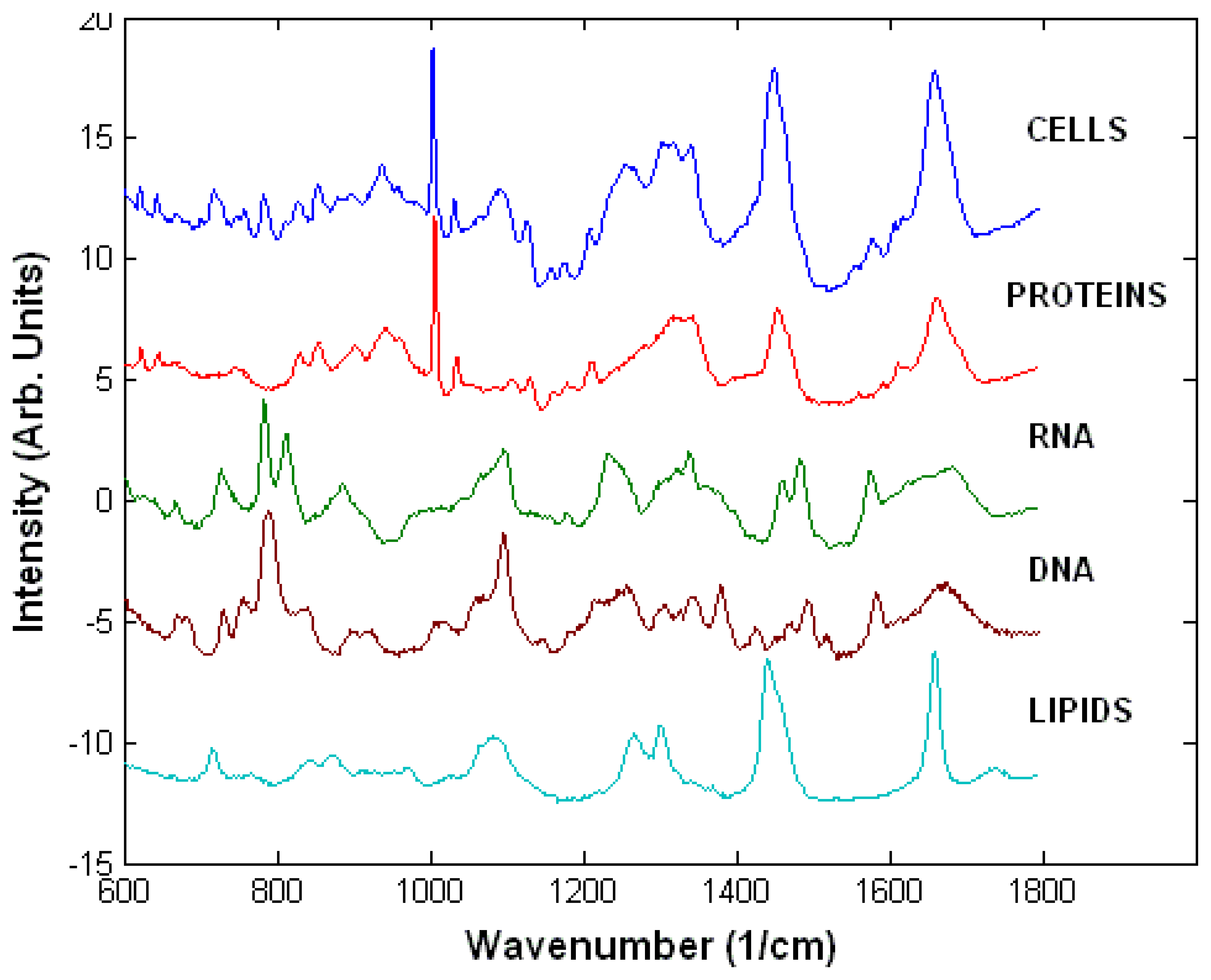
Raman spectrum-a cell's fingerprint. Raman spectrum of a single cell of...  | Download Scientific Diagram
Raman spectrum-a cell's fingerprint. Raman spectrum of a single cell of... | Download Scientific Diagram

Raman spectroscopy: an evolving technique for live cell studies - Analyst  (RSC Publishing) DOI:10.1039/C6AN00152A
Raman spectroscopy: an evolving technique for live cell studies - Analyst (RSC Publishing) DOI:10.1039/C6AN00152A

Example of an average Raman spectrum from a lung cancer cell (black)... |  Download Scientific Diagram
Example of an average Raman spectrum from a lung cancer cell (black)... | Download Scientific Diagram

In vivo lipidomics using single-cell Raman spectroscopy | PNAS

High Resolution Live Cell Raman Imaging Using Subcellular  Organelle-Targeting SERS-Sensitive Gold Nanoparticles with Highly Narrow  Intra-Nanogap | Nano Letters
High Resolution Live Cell Raman Imaging Using Subcellular Organelle-Targeting SERS-Sensitive Gold Nanoparticles with Highly Narrow Intra-Nanogap | Nano Letters
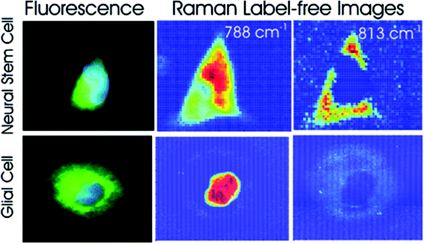
Using Raman Spectroscopy to Identify Cell Types | The Scientist Magazine®

Raman spectroscopy: an evolving technique for live cell studies - Analyst  (RSC Publishing) DOI:10.1039/C6AN00152A
Raman spectroscopy: an evolving technique for live cell studies - Analyst (RSC Publishing) DOI:10.1039/C6AN00152A

A) A schematic of the Raman cell assembly; (B) a schematic of the... |  Download Scientific Diagram
A) A schematic of the Raman cell assembly; (B) a schematic of the... | Download Scientific Diagram

Raman-Based in Situ Monitoring of Changes in Molecular Signatures during  Mitochondrially Mediated Apoptosis | ACS Omega
Raman-Based in Situ Monitoring of Changes in Molecular Signatures during Mitochondrially Mediated Apoptosis | ACS Omega

Average Raman spectrum of the secondary cell wall of poplar wood. The... |  Download Scientific Diagram
Average Raman spectrum of the secondary cell wall of poplar wood. The... | Download Scientific Diagram

Molecular imaging of live cells by Raman microscopy - ScienceDirect

Raman spectra of an individual cell. (a) Nucleus, (b) cytoplasm. | Download  Scientific Diagram
Raman spectra of an individual cell. (a) Nucleus, (b) cytoplasm. | Download Scientific Diagram